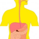
GASTROINTESTINAL MOTILITY
 The gastrointestinal tract is in a continuous contractile, absorptive, and secretory state. The control of this state is complex, with contributions by the muscle itself, local nerves (i.e., the enteric nervous system, ENS), the central nervous system (CNS), and humoral pathways. Of these, perhaps the most important regulator of physiological gut function is the ENS which is an autonomous collection of nerves within the wall of the GI tract, organized into two connected networks of neurons: the myenteric (Auerbach’s) plexus, found between the circular and longitudinal muscle layers, and the submucosal (Meissner’s) plexus, found below the epithelium. The former is responsible for motor control, while the latter regulates secretion, fluid transport, and vascular flow.
The ENS is responsible for the largely autonomous nature of most gastrointestinal activity. This activity is organized into relatively distinct programs that respond to input from the local environment of the gut and the CNS. Each program consists of a series of complex, but coordinated, patterns of secretion and movement that show regional and temporal variation. The fasting program of the gut is called the MMC (migrating myoelectric complex when referring to electrical activity and migrating motor complex when referring to the accompanying contractions) and consists of a series of four phasic activities.
The most characteristic, phase III, consists of clusters of rhythmic contractions that occupy short segments of the intestine for a period of 6 to 10 minutes before proceeding caudally. One whole MMC cycle (i.e., all four phases) takes about 80 to 110 minutes. The migrating motor complex occurs in the fasting state, during which it helps sweep debris caudad in the gut. The migrating motor complex cycles continually in animals that feed constantly, but is interrupted by another pattern of contractions¾the fed program¾in intermittently feeding animals such as humans. The fed program consists of high-frequency (12 to 15 per minute) contractions that are either propagated for short segments (propulsive) or are irregular and not propagated (mixing).
The basic motor tool used by the ENS to integrate its programs is the peristaltic reflex. Physiologically, peristalsis is a series of reflex responses to a bolus in the lumen of a given segment of the intestine; the ascending excitatory reflex results in contraction of the circular muscle on the oral side of the bolus, while the descending inhibitory reflex results in relaxation on the anal side. The net pressure gradient moves the bolus caudad. Three neural elements, responsible for sensory, relay, and effector functions, are required to produce these reflexes. Luminal factors stimulate sensory elements in the mucosa, leading to a coordinated pattern of muscle activity that is directly controlled by the motor neurons of the myenteric plexus to provide the effector component of the peristaltic reflex.
Motor neurons receive input from ascending and descending interneurons (which constitute the relay and programming systems) that are of two broad types, excitatory and inhibitory. The primary neurotransmitter of the excitatory motor neurons is acetylcholine (ACh), although tachykinins, co-released by these neurons, also play a role. The principal neurotransmitter in the inhibitory motor neurons appears to be nitric oxide (NO), although important contributions may also be made by ATP, vasoactive intestinal peptide, and pituitary adenylyl cyclase-activating peptide (PACAP), all of which are variably coexpressed with NO synthase.
This view of nerve-muscle interaction within the GI tract may be oversimplified, and other cell types may be important. One of these is the interstitial cell of Cajal, distributed within the gut wall and responsible for setting the electrical rhythm, and hence the pace of contractions, in various regions of the gut. These cells also may translate or modulate neuronal communication to the muscle, by mechanisms yet to be worked out.
Control of tension in gastrointestinal smooth muscle is in large part dependent on the intracellular Ca2+ concentration. In general, there are two types of excitation-contraction coupling. Ionotropic receptors can mediate changes in membrane potential, which in turn activate voltage-dependent Ca2+ channels to trigger an influx of Ca2+ (electromechanical coupling); metabotropic receptors activate various signal transduction pathways to release Ca2+ from intracellular stores (pharmacomechanical coupling). Inhibitory receptors also exist on smooth muscle and generally act via PKA and PKG, whose kinase activities can lead to hyperpolarization, decreased cytosolic [Ca2+], and reduced interaction of actin and myosin. As an example, NO may induce relaxation via activation of guanylyl cyclase, GMP-pathway, and the opening of several types of K+ channels.
Gastrointestinal motility disorders are a complex and heterogeneous group of syndromes whose pathophysiology is not completely understood. Typical motility disorders include achalasia of the esophagus (impaired relaxation of the lower esophageal sphincter associated with defective esophageal peristalsis that results in dysphagia and regurgitation), gastroparesis (delayed gastric emptying), myopathic and neuropathic forms of intestinal dysmotility, and others. These disorders can be congenital, idiopathic, or secondary to systemic diseases (e.g., diabetes mellitus or scleroderma). This term also has traditionally (and perhaps inaccurately) included disorders such as irritable bowel syndrome and noncardiac chest pain in which disturbances in pain processing or sensory function may be more important than any associated motor patterns. For most of these disorders, treatment remains empirical and symptom-based, reflecting our ignorance of the specific derangements in pathophysiology involved.
Direct activation of muscarinic receptors, such as with the older cholinomimetic agents is not a very effective strategy for treating GI motility disorders because these agents enhance contractions in a relatively uncoordinated fashion that produces little or no net propulsive activity. By contrast, prokinetic agents are medications that enhance coordinated GI motility and transit of material in the GI tract.
Although ACh, when released from primary motor neurons in the myenteric plexus, is the principal immediate mediator of muscle contractility, most of the clinically useful prokinetic agents act “upstream” of ACh, at receptor sites on the motor neuron itself, or even more indirectly, on neurons one or two orders removed from it. Although pharmacologically and chemically diverse, these agents appear to enhance the release of excitatory neurotransmitter at the nerve-muscle junction without interfering with the normal physiological pattern and rhythm of motility. Coordination of activity among the segments of the gut, necessary for propulsion of luminal contents, therefore is maintained.
By: Ammarah Khan


Do you mind if I quote a couple of your articles as long as I
provide credit and sources back to your weblog? My website is in the exact
same niche as yours and my visitors would certainly benefit from
some of the information you provide here. Please let me know if this ok with you.
Thank you!