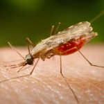
MALARIA
 Malaria especially that is caused by Plasmodium falciparum is the world’s most devastating human parasitic infection. Malaria afflicts nearly 500 million people and causes some 2 million deaths each year. Infection with P. falciparum, which preferentially affects children younger than 5 years of age, pregnant women, and nonimmune individuals, is responsible for nearly all this mortality. Although mosquito-transmitted malaria now is rare in North America, Europe, and Russia, its increasing prevalence in many other parts of the world poses a major international health and economic burden and a serious risk to travellers from nonendemic areas.
Inexpensive and safe drugs, insecticides, and ultimately, vaccines still are needed to combat malaria. In the 1950s, attempts to eradicate this scourge failed primarily because of the development of resistance to insecticides. Since 1960, transmission of malaria has risen in most regions where the infection is endemic, drug-resistant strains of P. falciparum have spread, and the degree of drug resistance has increased. More recently, chloroquine-resistant strains of P. vivax also have been documented.
Nearly all human malaria is caused by four species of obligate intracellular protozoa of the genus Plasmodium. Although malaria can be transmitted by transfusion of infected blood, congenitally, and by sharing needles, infection usually is transmitted by the bite of infected female Anopheles mosquitoes. Sporozoites from the mosquito salivary glands rapidly enter the circulation after a bite and localize via specific recognition events in hepatocytes, where they transform, multiply, and develop into tissue schizonts. This primary asymptomatic tissue (preerythrocytic or exoerythrocytic) stage of infection lasts for 5 to 15 days, depending on the Plasmodium species. Tissue schizonts then rupture each releasing thousands of merozoites that enter the circulation, invade erythrocytes, and initiate the erythrocytic cycle.
Once the tissue schizonts burst in P. falciparum and P. malariae infections, no forms of the parasite remain in the liver. However, in P. vivax and P. ovale infections, tissue parasites (hypnozoites) persist that can produce relapses of erythrocytic infection months to years after the primary attack. Once plasmodia enter the erythrocytic cycle, they cannot reinvade the liver; thus, there is no tissue stage of infection for malaria contracted by transfusion. In erythrocytes, most parasites undergo asexual development from young ring forms to trophozoites and finally to mature schizonts.
Schizont-containing erythrocytes rupture, each releasing 6 to 32 merozoites depending on the Plasmodium species. It is this process that produces febrile clinical attacks. The merozoites invade more erythrocytes to continue the cycle, which proceeds until death of the host or modulation by drugs or acquired partial immunity. The periodicity of parasitemia and febrile clinical manifestations depends on the timing of schizogony of a generation of erythrocytic parasites. For P. falciparum, P. vivax, and P. ovale, it takes about 48 hours to complete this process; for P. malariae, about 72 hours is required.
For erythrocyte invasion, merozoites bind to specific ligands on the red cell surface. P. falciparum has a family of binding proteins that can recognize a number of host cell molecules, including glycophorins A, B, and C, as well as band 3. It is able to invade all stages of erythrocytes and therefore can achieve high parasitemias. P. vivax is more selective in its binding; it needs to recognize the Duffy chemokine receptor protein as well as reticulocyte-specific proteins; thus, it will not establish infection in Duffy-negative individuals and will only invade reticulocytes. Because of this restricted subpopulation of suitable erythrocytes, P. vivax rarely exceeds 1% parasitemia in the bloodstream. P. ovale is similar to P. vivax in its predilection for young red blood cells, but the mechanism of its erythrocyte recognition is unknown. P. malariae recognizes only senescent red cells, maintains a very low parasitemia, and typically causes an indolent infection.
P. falciparum assembles cytoadherence proteins (the PFEMPs encoded by a highly variable family of var genes) into structures called knobs on the erythrocyte surface. This allows the parasitized erythrocyte to bind to the vascular endothelium, to avoid the spleen, and to grow in a lower oxygen environment. For the patient, the consequences are microvascular blockage in the brain and organ beds and local release of cytokines and direct vascular mediators such as nitric oxide, leading to cerebral malaria. It’s hard to capsulate everything about malaria in single article. Therefore, I’ll try to reveal some more facts in next article of mine.
By: Ammarah Khan


Woah! I’m really enjoying the template/theme of this blog. It’s simple, yet effective. A lot of times it’s tough to get that “perfect balance” between superb usability and visual appearance. I must say you’ve done a amazing job with this. In addition, the blog loads very quick for me on Safari. Exceptional Blog!
Thank You
I blog often and I truly appreciate your information. This great article has really peaked my interest. I’m going to book mark your website and keep checking for new details about once a week. I opted in for your RSS feed too.
Hmm it looks like your site ate my first comment (it was extremely long) so I guess I’ll just sum it up what I
submitted and say, I’m thoroughly enjoying your blog.
I too am an aspiring blog writer but I’m still new to everything.
Do you have any tips for inexperienced blog writers? I’d genuinely appreciate it.